Human Digestive System Diagram
Human digestive system diagram Starts From The ‘Mouth’ To The ‘ Anus ‘ ( Anus ). It Has The Following Parts- (I) Mouth ( Ii ) Oesophagous ( Iii) Stomach( Jv ) Small Intestine ( V) Large Intestine ( Vi) Rectum Digestion Takes Place In The Above Mentioned Organs.
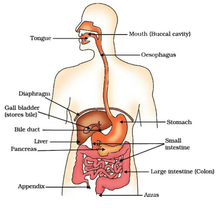
Human digestive system diagram – Mouth
In This , By Mixing With The Food From The Salivary Gland ( Saliva Gland ), It Gives Acidic Form To The Food And The Enzyme ‘ Amylase ‘ Or Starch Found In Saliva .
Partially Digested. The Taste Of Hot Food Increases In The Mouth Because The Surface Area Of The Tongue Increases. An Enzyme Found In The Mouth- ‘ Lysozyme ‘ Works To Kill Bacteria. Food Moves From The Mouth To The Front Of The Digestive System At The Rate Of Contractile Or Peristalsis.
Human digestive system diagram – Pharynx
No Digestion Takes Place In This Part. It Only Serves To Connect The Mouth And The Stomach .
Human digestive system stomach diagram
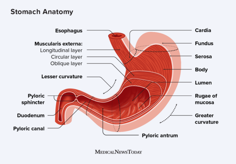
Digestion Of Food Takes Place In An Acidic Medium In The stomach diagram.
Gastric Glands Are Found In The Human Stomach, Which Secrete Gastric Juice.
The Maximum Amount Of Water Is Found In The Chemical Organization Of Gastric Juice.
Apart From This, HCl And Various Types Of Enzymes Are Found.
The Following Enzymes Are Found In The Stomach Whose Functions Are As Follows
(A) Pepsin Enzyme – Pepsin Enzyme : Digestive Of Protein Is Done By It.
(B) Renin Enzyme – Renin Enzyme : It Helps In Digestion Of Casein Protein Found In Milk.
(C) Lipase Enzyme – Lipase Enzyme : Digestion Of Fat By This Is – Digestive.
(D) Amylase Enzyme – Amylase Enzyme : It Helps In Digestion Of Starch. HCl Makes The Medium Of Digestion Of Food Acidic In The Stomach. It Kills Harmful Bacteria That Come With Food And Particles Like Pebbles And Stones.
Human digestive system diagram – Small Intestine
Digestion Of Food In The Small Intestine Takes Place In Alkaline Medium Because The PH Value Of Intestinal Juice Is 8.0 To 8.3.
The Small Intestine Is Considered The Longest Part Of The Alimentary Canal.
Whose Length Is About 6 To 7 Meters.
On The Basis Of Function And Structure, There Are Three Parts Of Small Intestine Which Are Called Duodenum, Midgut And Ileum Respectively.
Pittaras And Pancreatic Juices Are Helpful In The Digestion Of Food In The Duodenal Part Of The Small Intestine.
Formation Of Bile Juice In The Liver And Pancreatic Juice. The Formation Takes Place In The Pancreas.
Liver : Liver Is The Largest Exocrine Gland In The Human Body.
On The Basis Of Weight, The Liver Is Considered To Be The Largest Organ Of The Body, Weighing About 1500 Grams.
The Skin Is Considered The Largest Organ Of The Body On The Basis Of Length.
A Liver Is Found In Humans, Which Is Divided Into Two Bodies, In Which A Sac-Like Structure Is Found At The Bottom In The Right Body, Which Is Called Gall Bladder.
The Accumulation Of Bile Juice Takes Place In The Gall Bladder While The Formation Of Bile Takes Place In The Liver.
Gallbladder Is Not Found In Some Mammals. Like Horse, Zebra, Donkey, Mule And Mouse Etc.
The Bile Juice Produced In The Liver Is Alkaline In Nature With A PH Of About 7.7.
The Enzymes Are Not Found In Bile Juice, Yet Fat Is Digested By It Which Is Called Emulsification .
The Emulsification Process Is Related To The Liver.
Human digestive system diagram – Pancreas
The Pancreas Is Such An Organ Of The Human Body That Functions Like A Mixed Gland. Pancreatic Duct Is Found In The Pancreas As The Exocrine Part While The Isleit Of Langerhans Is Found As The Endocrine Part. Langerhans Islands Are Made Up Of Three Types Of Cells Called Alpha, Beta And Gamma Cells Respectively.
Alpha Cells : Alpha Cells Secrete The Hormone Glucagon. This Hormone Increases The Amount Of Glucose In The Blood.
Beta Cells : These Cells Secrete The Hormone Insulin Which Controls The Amount Of Glucose In The Blood.
Insulin – Hyposecretion Of The Hormone Insulin Increases The Amount Of Glucose In The Blood, Which Is Called Diabetes Mellitus .
Pancreatic Juice Is Produced In The Pancreas, Which Is Known As Complete Digestive Juice Because It Contains Enzymes That Fully Digest All Types Of Nutrients.
For Example, Trypsin Enzyme Is Found For The Digestion Of Proteins.
The Intestinal Glands Found In The Small Intestine Are Called Brunner’s Glands.
In Which Intestinal Juice Is Formed.
In Which Enzymes That Fully Digest All Types Of Nutrients Are Found, Which Digest Carbohydrates In This Way ( Sucrase Enzyme ): By This The Digestion Of Sucrose Sugar Takes Place.
Lactase Enzyme – Lactase Enzyme : It Helps In Digestion Of Lactose Sugar Found In Milk. Maltase Enzyme: It Helps In Digestion Of Maltose Sugar Found In Seeds.
Protein Digestive Enzyme Erepsin Enzyme – Erepsin Enzyme : Complete Digestion Of Protein Takes Place By This. That Is, This Enzyme Breaks Down Proteins Into Amino Acids.
Fat Digestive Enzyme: Lipase Enzyme: It Helps In Digestion Of Fats Into Fatty Acids And Glycerol.
Human digestive system diagram – Large Intestine
In This Part, The Remaining Food Is Absorbed And The Remaining 90% Water Is Absorbed.
The Length Of The Large Intestine Is 1 To 1.5 Meters, Where Digestion Of Food Does Not Take Place.
Basis Of Function And Structure There Are Three Parts Of The Large Intestine Which Are Respectively Called Esophagus, Colon And Rectum.
Human digestive system diagram – Rectum
Residual Food Is Stored In This Part.
From Here The Exodus Takes Place From Time To Time.
Note- Cellulose (A Type Of Complex Carbohydrate) Is Not Digested In Our Body.
Digestion Of Cellulose Takes Place In The Cecum . ‘Cecum’ Is Found In Herbivorous Animals.
The Cecum Is Left As A Dormant Organ In Humans.
The Structure Of The Tubule Attached To The Ceacum Is Called The Vermiform Appendix, Which Is A Residual Structure In Humans, That Is, At Present There Is No Function Of This Structure In The Human Body.
In Herbivorous Animals, The Worm Form Sphincter Helps In The Digestion Of Cellulose.
This Structure Is Not Found In Carnivorous Animals. Appendicitis Is Caused By The Increase Of The Worm Form.
Lorem Ipsum is simply dummy text of the printing and typesetting industry. Lorem Ipsum has been the industry’s standard dummy text ever since the 1500s, when an unknown printer took a galley of type and scrambled it to make a type specimen book.


